Near-infrared spectroscopy (NIRS) shows tremendous promise for a range of research and clinical applications. Because it is noninvasive and portable, and uses safe non-ionizing light, it allows uses that are impracticable or simply not possible with modalities such as MRI and positon emission tomography. And because the technology is relatively inexpensive, a much broader base of users can take advantage of its robust functionality.
The Theory
But what is near-infrared spectroscopy? A simple experiment sheds light on this question.
If you shine a flashlight onto your hand, you will see that the light can still be detected after traveling through several centimeters of tissue. Having observed this, you might ask yourself: If light can be recovered after passing through the body, can it be used to see, or image, inside the body? Advances in our understanding of light migration through tissue, the resulting development of tomography algorithms, and subsequent experimental verification have shown that imaging with diffuse light - using near-infrared spectroscopy and diffuse optical imaging (DOI) - is in fact possible.
Optical imaging at centimeter depths is afforded by the relationship of the absorption spectra of water, oxygenated hemoglobin (HbO) and deoxygenated hemoglobin (Hb), the three primary absorbers in tissue at near-infrared wavelengths. The water spectrum at those wavelengths permits a "spectral window” in the background absorption allowing investigators to see the hemoglobin. Moreover, within this window, the spectra of oxygenated and deoxygenated hemoglobin are distinct enough to allow spectroscopy and recovery of separate concentrations of both types of molecules. The scattering properties of different tissue types are similarly distinct.
Using diffuse optical imaging, researchers can then reconstruct the three-dimensional spatial variations in blood parameters: including hemoglobin concentration and oxygen saturation as well as tissue scattering characteristics. Thus, using light, they can effectively "see" inside the body.
What Does The Data Look Like?
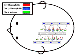
NIRS data consists of a series of time-dependent signals measured between individual light source and detector positions on a probe. The concentrations of oxygenated, deoxygenated and total hemoglobin can be calculated for each source-detector pair. These time-courses may represent, for example, the subject’s averaged hemodynamic response to repeated stimuli or tasks such as sensory stimuli or motor responses.
The data is often displayed topographically according to the positions of sources and detectors used. Probes are often designed for study of a specific region of the brain. We can develop custom probes to meet your specific needs.
Learn NIRS With Hands-On Training
To learn more about NIRS technology, TechEn recommends attending a course offered by Dr. David Boas at the MGH-Martinos Center for Biomedical Imaging. The introductory course covers the fundamentals of this optical technique and offers hands-on experience in its application.
Details about the course are available here.
Applications
NIRS can be applied in a number of areas. For example, since the late 1990s, increasing numbers of researchers have used it for brain mapping studies. They have employed visual, auditory and somatosensory stimuli to identify areas of the brain associated with certain cognitive functions; other areas of investigation have included the motor system and language. The technique could also contribute to the diagnosis and treatment of depression, schizophrenia and Alzheimer's disease. Researchers are already using it to understand the pathogenesis of these and a variety of other psychiatric disorders.
NIRS also shows promise as a clinical research tool, especially as it relates to the brain. Researchers are using the method to address the prevention and treatment of seizures and psychiatric concerns such as depression, Alzheimer’s disease and schizophrenia, as well as stroke rehabilitation. Furthermore, several groups are working to introduce the technology into the clinic itself - for instance, for neonatal monitoring and breast cancer detection.
-
Multi-modal Simultaneous Data Acquisition
Several applications take advantage of the multi-modal possibilities of NIRS. The optical technique can be integrated with MRI, EEG and trans-cranial magnetic stimulation (TMS) to provide colocalized, complementary information from each of the modalities. With MRI, for example, it can offer functional information about about brain hemodynamics while MRI provides structural information with which to localize the changes. Multi-modal imaging opens up a number of new opportunities for NIRS, and represents one of the most exciting areas of growth for the technique.


